
About our services
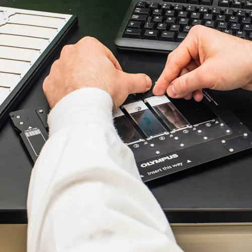
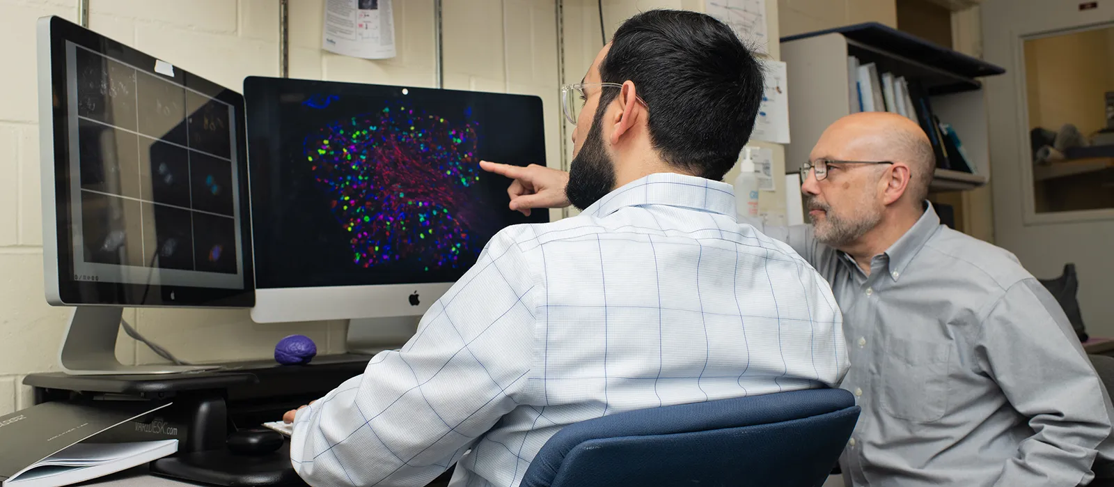
The Histology and Imaging Core provides investigators, both faculty and students, with access to specialized instrumentation and training related to tissue processing, sectioning, staining, immunohistochemistry, and microscopy.
Request Services
Work and training service requests are made via iLab.
Complete a work or training request
View instructions on setting up an iLab account (PDF)
Equipment
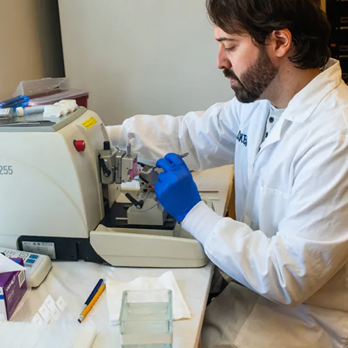
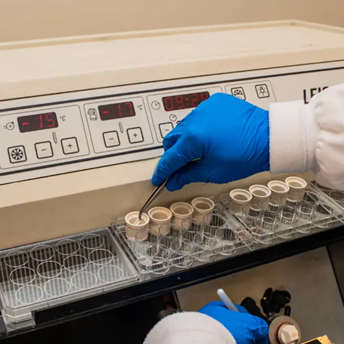
Instrumentation includes tissue embedding equipment, microtomes, vibratomes, cryostats, and slide-staining workstations.
- Leica RM2265 fully automated rotary microtome
- Sakura TEC 5 embedding center
- Sakura VIP E150 automated tissue processor
- Leica VT1000S vibrating microtome
- Leica CM1950 and CM1900 cryostats
- Biocare Medical Decloaking chamber for heat-induced epitope retrieval
- LifeCanvas SmartBatch electrophoretic tissue clearing and antibody labelling instrument
Technical Services
Technical services include paraffin, plastic and frozen embedding and sectioning of routine tissues and undecalcified bone; tissue clearing; special stains; immunohistochemistry; image acquisition and analysis, and antibody optimization and validation.
Sample Processing
The Histology and Imaging Core is fully integrated with the UNE’s Behavior Core and can directly process samples collected by the Behavior Core.