Biddeford Art Gallery presents 'Images of Neuroscience, Images of Transcendence' opening May 5
The Art Gallery at the University of New England’s Jack S. Ketchum Library in Biddeford is pleased to present “Images of Neuroscience, Images of Transcendence: Work from UNE’s Histology and Imaging Core and Paintings by Honour Mack,” opening May 5.
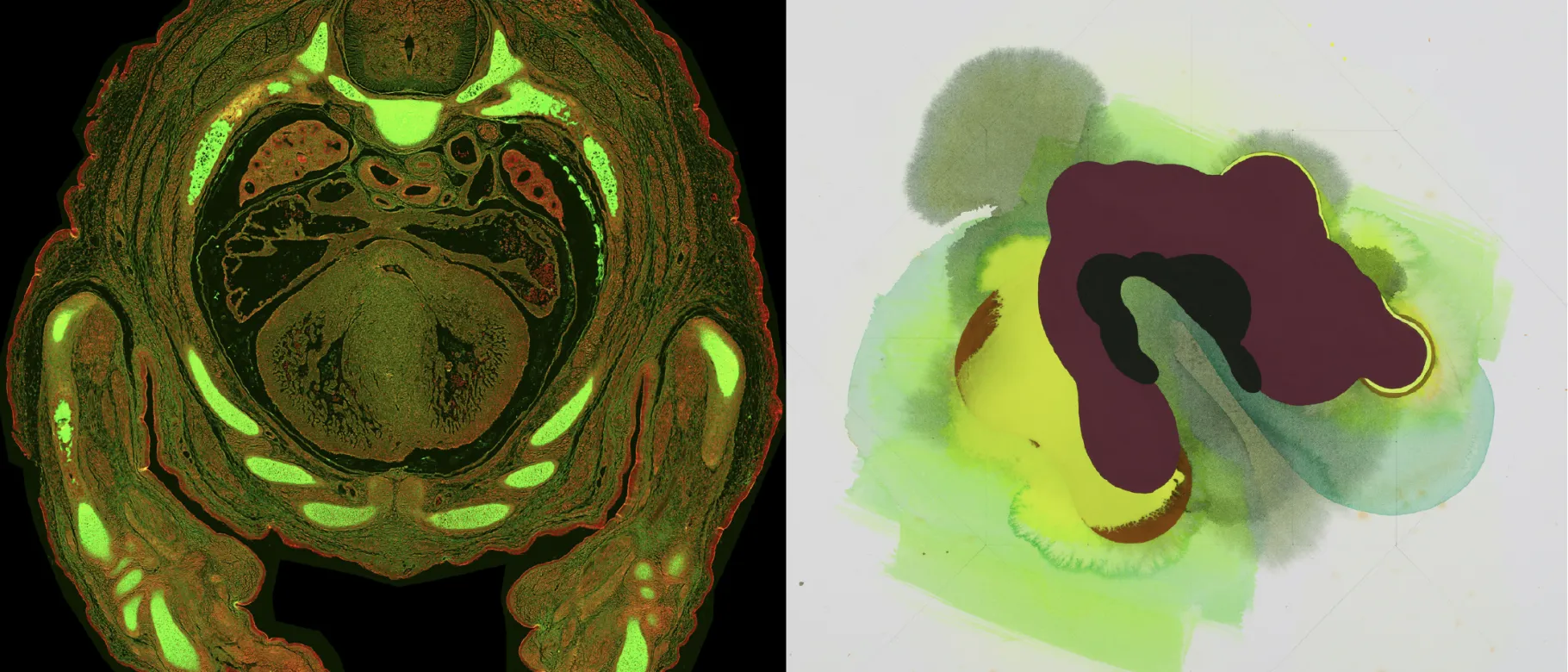
The exhibit will feature images of biological samples produced by scientists on UNE’s Biddeford Campus in conversation with the work of contemporary Maine painter Honour Mack.
“The energy of Mack’s paintings — colorful, geometrical, spiritual, and biomorphic — form a wonderful pairing with the striking images used for scientific inquiry, allowing a visual conversation to emerge from two different but sympathetic sources,” remarked Hilary Irons, director of Galleries and Exhibitions at UNE.
The exhibit is coordinated in collaboration with UNE’s Center for Excellence in the Neurosciences.
“My paintings offer no concrete destination but seek to suspend a viewer in the present moment,” Mack remarked, offering a unique counterpoint to the precision that science can provide. “This objective is supported by my interest in the intersection of references to anatomy, man-made structures, early esoteric philosophies, and ancient scientific examinations of the world.”
“Images of Neuroscience, Images of Transcendence” runs through Sept. 29. An opening reception for the exhibit will be held Friday, May 5, from 4 to 6 p.m. at the library, located at 11 Hills Beach Road, Biddeford. The library is open 9 a.m. to 5 p.m. daily.
For more information, contact Irons at hirons@une.edu.
Masks are optional indoors on UNE’s campuses. For UNE’s latest COVID-19 protocols, visit the UNE Onward website. Irons would like to extend special thanks to Ian Meng, Ph.D., director of UNE’s Center of Biomedical Research Excellence (COBRE) for the Study of Pain and Sensory Function, and Peter Caradonna, B.S., manager of COBRE’s Histology and Imaging Core.
Above images: (L-R): Mouse embryo transverse section stained with alcian blue and imaged with brightfield microscopy (image psuedocolored). Section prepared by Peter Caradonna, manager, COBRE Histology and Imaging Core at UNE, and Dr. Lindsey Fitzsimons, former Ph.D. student in the Tucker Lab at UNE; Honour Mack, Swank Bight, 2020, watermedia on paper, 22”x 22.”